
Gut Microbes, December 2025 · PMID 41376334 · animal evidence
Ewingella americana in the 2025 antitumour experiment: what was measured, panel by panel
Forty-five strains out of three animals, nine survivors, one that emptied the tumour. The study in the detail the headlines dropped — including the parts the authors themselves say are not settled.
Iwata, Yamasita, Asukabe, Sakari and Miyako, at the Japan Advanced Institute of Science and Technology in Nomi, Ishikawa, isolated 45 bacterial strains from the intestines of a Japanese tree frog, a fire-belly newt and a grass lizard; carried nine through biocompatibility screening; and found that one — Ewingella americana, from the frog — eliminated established Colon-26 tumours in every BALB/c mouse that received a single intravenous dose. Published in Gut Microbes 2025;17(1):2599562, PMID 41376334, PMC12710904. Animal evidence
1. The design
The premise first, because it is the unusual part: the authors went looking in animals nobody screens.
The gut microbiome of mammals has been mined hard for therapeutic organisms. The microbiomes of lower vertebrates have not. The team’s starting position was that amphibians and reptiles — animals living in environments dense with microbial competition — might carry species with properties no mammalian screen would surface.
- Sources Dryophytes japonicus (Japanese tree frog), Cynops pyrrhogaster (fire-belly newt), Takydromus tachydromoides (grass lizard)
- Isolated 45 distinct strains
- Carried forward 9, after biocompatibility screening
- Of those 9 six were Ewingella americana, all from the frog
- Tumour model syngeneic Colon-26 murine colon carcinoma
- Host female BALB/c mice
- Inoculum 1 × 10⁶ cells, subcutaneous, in 100 µL
Comparators were run in the same study rather than quoted from the literature, which is the reason the comparison carries weight: an anti-PD-L1 checkpoint antibody and liposomal doxorubicin, each at 2.5 mg/kg intravenously, every other day, four doses. The bacterium got one dose, once.
2. Efficacy
| Arm | Schedule | Outcome at day 30 |
|---|---|---|
| Ewingella americana | one IV dose, 200 µL of 5 × 10⁹ CFU/mL | 100% complete response; 100% survival |
| Anti-PD-L1 antibody | 2.5 mg/kg IV ×4, alternate days | one complete response |
| Liposomal doxorubicin | 2.5 mg/kg IV ×4, alternate days | no consistent tumour eradication |
| Saline control | — | uniform tumour growth |
3. Where the bacteria went
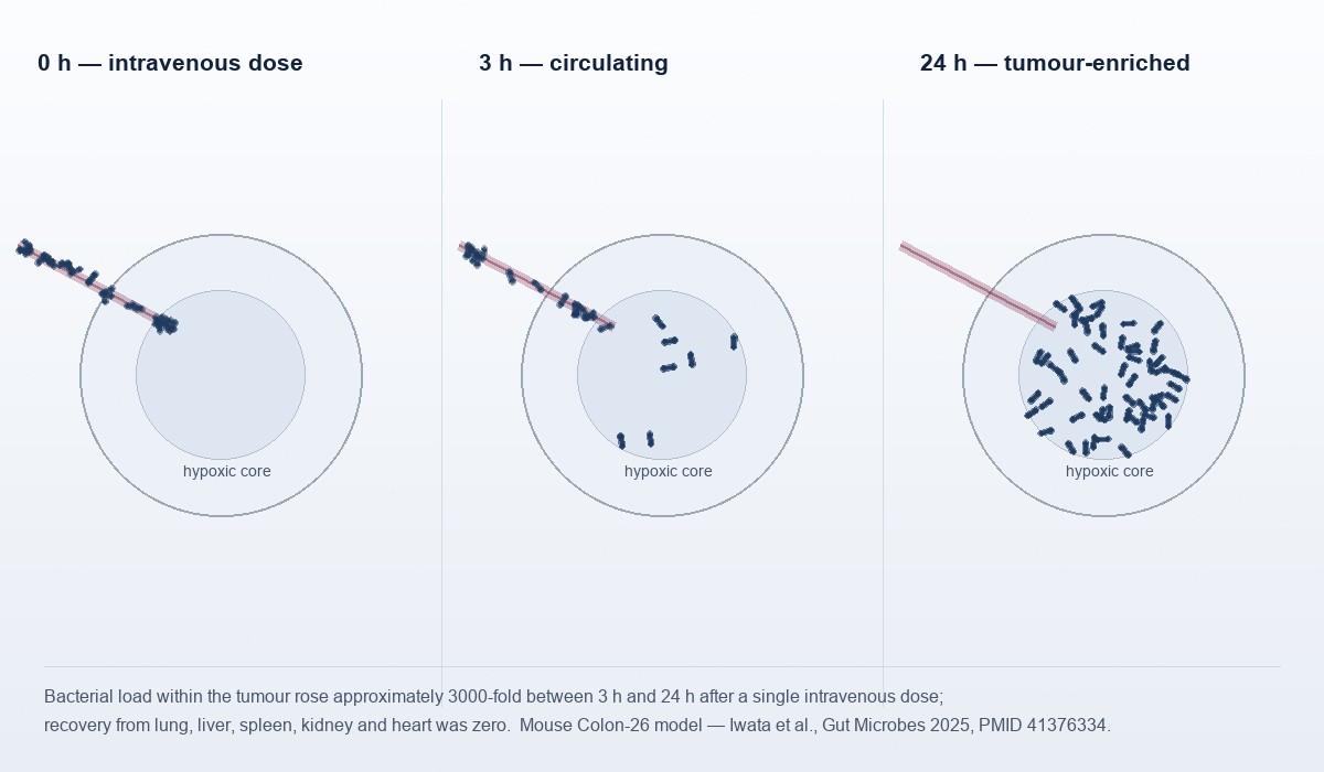
Biodistribution was the part of the study that turned an interesting cytotoxicity result into a plausible therapeutic mechanism. Bacterial load within the tumour rose approximately 3000-fold between 3 and 24 hours after the single intravenous dose. Recovery from lung, liver, spleen, kidney and heart was zero. Blood colony counts fell to undetectable by 24 hours.
The authors describe the result as tumour-exclusive localisation and attribute it to the intrinsic properties of a facultative anaerobe in hypoxic tissue — while stating explicitly that hypoxia is necessary but insufficient, since other hypoxic tissue was not colonised. That honesty is the most useful sentence in the paper: it says the mechanism of selectivity is not yet understood.
4. The dual mechanism
Direct killing
Secreted cytolysins — haemolysin and exotoxin activity. In three-dimensional tumour spheroid culture, the highest tested concentration (5 × 10⁸ CFU) largely destroyed the spheroid within 24 hours. In-vitro evidence
Virulence factors were identified computationally from the published genome of the type strain ATCC 33852 — not from the amphibian isolate itself, which is a real gap in the mechanistic chain.
Immune activation
Within treated tumours: CXCR4+ neutrophils rose 30%, CD3+ T cells 5%, CD19+ B cells 3%. Interferon-γ and TNF-α were elevated in tumour tissue.
A large live bacterial population inside an immunologically quiet tumour is an alarm that cannot be ignored. The rechallenge result says the alarm produced memory.
5. Safety, as measured in this study
Haematological and biochemical parameters showed no significant difference from saline-treated controls. Histology of liver, spleen, heart, lungs and kidneys found no bacterial presence, tissue damage, inflammatory infiltration or necrosis. Body weight did not differ significantly between groups by day 15. Blood clearance was complete by 24 hours; the authors describe only transient, mild responses resolving within 240 hours.
6. The authors’ own limitations
- A subcutaneous model, not orthotopic — future orthotopic studies named as needed
- One cancer type; broad-spectrum activity not established
- Dose fractionation flagged as a consideration for clinical translation
- Selectivity mechanism incompletely explained — hypoxia necessary, not sufficient
To which the honest reader adds: one laboratory, and no independent replication yet. Human interventional evidence: none
The complete evidence ledger, tier by tier → · Where this sits in a century of bacterial cancer therapy →
7. Trending in the field
8. References
- Iwata S, Yamasita N, Asukabe K, Sakari M, Miyako E. Discovery and characterization of antitumor gut microbiota from amphibians and reptiles. Gut Microbes 2025;17(1):2599562. PMID 41376334. PMC12710904. PubMed record
- Jafarova Ayik H, Eyupler C, Yassa G, Aksu C, Duman N. Sepsis caused by Ewingella americana in an immunocompromised patient. Cureus 2025;17(6):e85641. PMID 40636648. PubMed record
- Liu Z, Sheng H, Okorley BA, Li Y, Sossah FL. Comparative genomic analysis of the genus Ewingella. Pathogens 2020;9(5):330. PMID 32354059. PubMed record



